|
Sterile Cornea Infiltrates Related to Soft Contact Lens WearRen├® G. M├®ly, MDPresented in part at the CLAO Annual Meeting 2001 in Las Vegas
Abstract
Purpose: To evaluate the prevalence and associated risk factors of sterile cornea infiltrates in a population of contact lens wearers. Methods: A retrospective chart review of all cases seen by the author was conducted for unscheduled visits related to contact lens wear over a period of five years (1996-2000). Results: Among the one hundred and twenty-nine patients seen for unscheduled contact lens related visits, forty-one (31.78 %) had cornea infiltrates. Only one case presenting an anterior chamber reaction was suspected to be infectious, all other forty cases were sterile. All patients with infiltrates wore soft lenses. Nine patients (21.95 %) wore conventional soft lenses and thirty-two (78.05 %) wore frequent replacement lenses. None of the patients required hospitalisation and all cases were resolved after topical treatment. Among the forty sterile infiltrates, nine patients (6.97 %) had contact lens induced peripheral ulcers (CLPU) and thirty one had subepithelial infiltrates giving an estimated prevalence of 6.97% and 24.03 %, respectively, for all unscheduled contact lens related visits. Thirty-seven patients (92.5%) from this group used, presumably, multi-purpose disinfecting solutions and only three patients (7.5%) used a one-step 3% hydrogen peroxide system. Conclusions: Sterile cornea infiltrates are a frequent complication related to soft contact lens wear. Our major concern is to differentiate these innocuous lesions from infectious ulcerative keratitis. It is likely that contact lens hygiene plays a key role in the pathophysiology of sterile infiltrative events and patients should be better educated regarding contact lens care. Nevertheless, larger prospective studies are required to establish specific risk factors.
Key Words: Sterile Cornea infiltrates ŌĆō Contact lenses ŌĆō Keratitis ŌĆō Contact lens induced peripheral ulcers ŌĆō Subepithelial cornea infiltrates - Contact lens induced acute red eye ŌĆō Microbial keratitis - Keratitis nummularis ŌĆō Acanthamoeba keratitis ŌĆō Viral keratitis ŌĆō Fungal keratitis ŌĆō Contact lens hygiene
Introduction
Sterile corneal infiltrates related to contact lens wear were first reported twenty-five years ago by Bernstein and co-workers1. In a recent study, Cutter2 found a 15.1% prevalence of cornea infiltrates among one hundred and seventy-nine patients seen for unscheduled visits. The most important issue is to differentiate these common and innocuous lesions from infectious keratitis, which is still a potentially blinding condition.
The most common symptoms related to sterile infiltrates are redness, foreign body sensation, discomfort rather than pain, photophobia and lacrimation. Furthermore, there is no purulent discharge2,3,4 . In some cases of central infiltrates, patients complain of a slight blurring of their vision. Sterile corneal infiltrates may also be asymptomatic.
Sterile cornea infiltrates include a wide spectrum of clinical findings. Based on localisation and status of the epithelium, sterile infiltrates may be subclassified as contact lens induced peripheral ulcers (CLPU), and epithelial or subepithelial infiltrates.
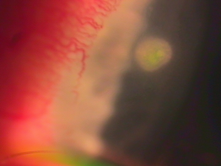
Contact lens induced peripheral ulcers (CLPU) are a distinct clinical entity. Grant and co-workers5 define CLPU as usually single, small (<2 mm diameter) lesions in the cornea mid-periphery or periphery with a full thickness epithelial defect and fluorescein penetration to the stroma at early stages (Figure 1); thus, indicating that the anterior layers of the stroma are involved. CLPU bear some resemblance to that of non-infectious hypersensitivity reactions to Staphylococcus aureus6. As with most authors, Grant5 found CLPU to be more common with extended wear of hydrogel lenses. The lesions are self-limiting and heal after removal of the lens within a few days - even without any medical treatment. Small grey scars are left and these can persist for months.
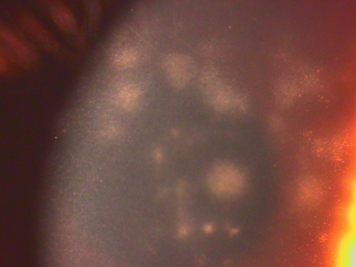
Fig. 1: CLPU in the cornea periphery with epithelial defect and fluorescein penetration to the stroma Sterile cornea infiltrates may also appear as epithelial or subepithelial grey-colored opacities less than 1.5 mm in diameter with an intact epithelium or mild punctate staining. They are usually multiple, midperipheral or centrally located (Figure 2). In many cases they appear as nummular opacities resembling that of adenoviral infections. They are often observed in association with contact lens related acute red eye (CLARE), especially after overnight wear of low Dk soft lenses7.
Figure 2: Subepithelial cornea infiltrates related to soft contact lens wear The differential diagnosis of cornea infiltrates includes the following: bacterial keratitis, fungal keratitis, Acanthamoeba keratitis and viral keratitis, and can usually be done on clinical grounds. Many studies have been performed to differentiate the relatively innocuous non-infectious sterile infiltrates from sight threatening bacterial keratitis. Most authors found that the presence of significant pain, purulent discharge, or anterior chamber reaction was associated with an infectious keratitis3,5,6. The status of the overlying epithelium is subject to controversy4. Staining lesions are more likely to be infectious3, but some cases of infection without any epithelium defect have been reported8. On the other hand lesions such as CPLU are sterile, although associated with a full thickness epithelial defect at early stages5. Multiple infiltrates are more likely to be sterile whereas the presence of corneal oedema around an infiltrate is indicative of infection9.
Fungal cornea infections are indolent and have grey infiltrates, often with satellite lesions. Marked ocular inflammation and hypopyon are features that distinguish this condition from sterile infiltrates.
Acanthamoeba is a free-living protozoan that thrives in polluted water. The initial symptoms of Acanthamoeba keratitis are pain out of proportion to the clinical findings. At early stages, slit lamp examination may reveal granular epithelial irregularity with punctate or dendritiform changes followed by ring infiltrates, perineural infiltrates and anterior uveitis8 . Acanthamoeba keratitis is differentiated from sterile infiltrates by the presence of significant pain with mild signs of inflammation10.
Nummular cornea opacities are most common in epidemic keratoconjunctivitis (EKC) caused by adenoviruses 8 and 19, but may also be seen in other virus infections such as adenoviruses 3, 4 and 7, herpes zoster and Epstein-Barr. In EKC, the subepithelial lesions appear 8-15 days after onset of conjunctivitis and may persist for months or even years. A history of preauricular node, oedema of the eyelids, subconjunctival haemorrhages and chemosis eliminates sterile contact lens related infiltrates11.
Extended wear of hydrogel lenses, poor lens hygiene, microbial contamination of lens cases, hypoxia and dry eye conditions are well established risk factors leading to corneal infiltrative events6,2,12,13.
The term ŌĆ£sterile infiltratesŌĆØ is in some ways confusing. In a number of studies, cultures taken from presumed sterile infiltrates yielded bacteria8,10. Microbial contamination of lens cases is also very common in patients presenting CLARE or cornea infiltrates6,13. Yet, the types of bacteria found in patients with sterile infiltrates include less virulent microorganisms and are therefore different from severe cases of bacterial keratitis where Pseudomonas is predominant. Nevertheless the question, whether the presence of bacteria represents a true infection or is related to a hypersensitivity response, remains controversial13.
Although the pathophysiology of corneal infiltrates is not completely understood, it is likely that it is an immunologically mediated reaction6. In an experimental study performed on rabbits by Mondino15, animals immunised by intradermal or subconjunctival injections of Staphylococcus aureus antigens followed by topical application of viable bacteria developed corneal phlyctenules and catarrhal infiltrates.
In contact lens wearers many agents may present an antigenic load as follows:
- Lens material itself1 - Lens care solutions and preservatives such as thimerosal16 - Deposits - Bacteria or toxins adherent to the lens13
There is some evidence that sterile cornea infiltrates are healing after lens removal without any medical treatment5. However, most authors recommend treating presumed sterile cornea infiltrates with appropriate topical antibiotics for a few days rather than risk a potentially devastating complication. Patching is contraindicated. The patient should be re-examined the next day. If inflammation is persisting after a few days without any signs of infection, topical corticosteroids may be added to the initial antibiotic treatment4,9. After the lesions have healed, patients should use new lenses and new lens cases. A careful history evaluating compliance and lens care is most important. This is especially true in cases of epithelial infiltrates, and preservative-free care products should be recommended. Patients who previously wore their lenses on an extended wear basis should change to a daily wear modality4,9.
Methods and Materials
A retrospective chart review of all cases seen in my ophthalmological practice was conducted for unscheduled visits related to contact lens wear over a period of five years (1996-2000).
The following information was collected from the charts: - Age and gender - Type of lens worn and wear modality - Lens care systems used - Type and location of the infiltrate - Status of the anterior chamber - Possible risk factors
Infiltrates were considered to be peripheral if located within 2 mm from the limbus; others were described as central.
Culture results were not included in the study.
Results
Among the seven hundred and twenty-one contact lens wearers examined in my office over the past five years, one hundred and twenty-nine came for unscheduled visits related to various contact lens problems. This gave an estimated prevalence of 17.64%.
Lens type and modality: The large majority of these one hundred and twenty-nine patients wore soft lenses (113 patients, 87.59 %). Fifteen patients (11.62%) wore rigid gas permeable (RGP) lenses and one child (0.77%) wore silicone lenses (Silflex). Only four patients wore their lenses on an extended wear basis. For two of the cases this was because of aphakia and in the other two as a bandage lens. Among the one hundred and thirteen patients wearing soft lenses, twenty-eight (24.77%) wore conventional soft lenses and eighty-five (75.22 %) wore frequent replacement lenses.
Prevalence of infiltrates: Among these one hundred and twenty-ninepatients, forty-one had cornea infiltrates giving an estimated prevalence of 31.78 % of all unscheduled contact lens related visits and 5.6% of the contact lens population of my practice. Only one case presenting an anterior chamber reaction was suspected to be infectious, whereas all other forty cases were sterile.
Characteristics of the population having infiltrates: Among the forty-one patients having infiltrates only fourteen (34.14 %) had been fitted in my practice, the other twenty-seven patients were coming for the first time. The mean age of this group was twenty-nine years. Nearly two-thirds of the patients were female (63.41%) and slightly more than one-third were males (36.58%). Nine patients (21.95 %) wore conventional soft lenses and thirty-two (78.05 %) wore frequent replacement lenses.
Population having CLPU: Nine patients presented CPLUs giving an estimated prevalence of 6.97 % of all unscheduled contact lens related visits and 1.23 % of the contact lens population of my practice. Only two patients from this group wore lenses on an extended wear basis: One elderly lady wore a conventional soft extended wear lens for unilateral aphakia, and one young man wore a silicone hydrogel lens because of lagophthalmos. Eight patients had a solitary ulcer; only one patient presented two CLPUs on the same eye.
Population having subepithelial infiltrates: Thirty-one patients had subepithelial infiltrates giving an estimated prevalence of 24.03 % of all unscheduled contact lens related visits and 4.24 % of the contact lens population of my practice. None of these patients wore extended wear lenses but one patient reported having slept in his lenses. In sixteen cases, the infiltrates were located on both eyes, in seven cases on the right eye and in eight cases on the left eye.
Lens care regimen: Among the thirty-eight patients presenting sterile corneal infiltrates and wearing daily wear lenses, only three (7.9%) used a one-step 3% hydrogen peroxide care system while twenty-nine patients used various multi-purpose disinfecting solutions. Six patients used, presumably, multi-purpose disinfecting solutions but could not name the brand. This gave an overall presumed prevalence of multi-purpose solutions of 92.1%. Unfortunately, the number of patients in each subgroup of specific products was too small for a valuable statistical analysis. Two patients wore extended wear lenses and used no care products. Discussion
Since most studies are conducted in eye hospitals, the literature contains little data describing minor complications related to contact lenses in general ophthalmological practices. Due to the design of this retrospective study the overall incidence of infiltrative events could not be determined. Forty cases of sterile infiltrates were identified among the one hundred and twenty-nine patients seen for contact lens related unscheduled visits in my practice giving a prevalence of 31.5% for this group of patients. This is much higher than in the study of Cutter2 and co-workers who found a prevalence of 15.1% among one hundred and seventy-nine patients seen for unscheduled visits. This may be related to the fact that only patients presenting an overlying staining were considered in this study whereas all infiltrates, including minor infiltrative events without staining, have been included in my study. Overnight wear is still very uncommon in Germany and plays no significant part in the aetiology of infiltrates in this study. Nevertheless many people occasionally sleep in their lenses and among the group of one hundred and twenty-nine unscheduled visits, fourteen cases (10.85%) of CLARE were related to overnight wear of low Dk hydrogels. Only one patient among this group had associated cornea infiltrates. The incidence of severe keratitis was found to be significantly lower for daily wear of disposable lenses than for daily wear of conventional soft and RGP lenses in two extended Swedish studies17. Nevertheless, I found a high percentage (78.05%) of frequent replacement lenses in the infiltrative group of my study and this had also been noticed in the study of Cutter2 and co-workers. An other interesting finding was the very high percentage (92.1%) of multi-purpose solutions used in this group. At first glance, the large number of infiltrative complications in my study could be related to these two major findings. As confirmed in a recent study13, it is well established that bacterial contamination of lenses and lens cases can induce CLARE responses and cornea infiltrates. Although most multi-purpose solutions have been proven in tests to provide a sufficient antimicrobial activity, contaminated lens cases are very common findings in the literature. The problem seems not to be the type of lens or the product used for lens care, but simply the way in which they are used. An increasing number of frequent replacement lenses seems not to be fitted by professionals. Internet sales of replacement lenses are rapidly growing. As a result, frequent replacement lens wearers are likely to be less educated regarding lens hygiene as wearers of conventional wearers used to be. Many people using multi-purpose solutions do not follow the recommendations made by manufacturers and keep the same solution in their lens cases for weeks. This phenomenon is probably seldom in case of people using one or two step hydrogen peroxide systems. Deposits on frequent replacement lenses are common findings indicating that the planned replacement schedule is not always respected. Unfortunately my study cannot evaluate the causal effect of these factors: that will require larger prospective studies in order to perform statistic analyses of each hypothesis.
ConclusionSterile cornea infiltrates are a frequent complication related to soft contact lens wear. Our major concern is to differentiate these innocuous lesions from infectious ulcerative keratitis. It is likely that contact lens hygiene plays a key role in the pathophysiology of sterile infiltrative events and patients should be better educated regarding compliance and contact lens care. Nevertheless, larger prospective studies are required to establish specific risk factors.
References
1. Bernstein HN, Kemp MA: An unusual keratoconjunctivitis occurring after longtime wearing of the AO softcon (formerly Griffin or Bionite) hydrophilic contact lens. Ann Ophthalmol 1975;7:87-106. 2. Cutter GR, Chalmers RL, Roseman M: The clinical presentation, prevalence, and risk factors of focal corneal infiltrates in soft contact lens wearers. CLAO J 1996;22:30-37. 3. Stein RM, Clinch TE, Cohen EJ, et al: Infected vs. sterile cornea infiltrates in contact lens wearers. Am J Ophthalmol 1988;106:632-636. 4. Donshik PC: Peripheral corneal infiltrates and contact lens wear. CLAO J 1998;24:134-135. 5. Grant T, Chong MS, Vajdic C, Swarbrick HA, Gauthier C, Sweeney DF, Holden BA: Contact lens induced peripheral ulcers during hydrogel contact lens wear. CLAO J 1998;24:145-151 6. Bates AK, Morris RJ, Stapleton F, Minassian DC, Dart JKD: ŌĆ£SterileŌĆØ corneal infiltrates in contact lens wearers. Eye1989;3:803-810. 7. Holden BA, La Hood D, Grant T, Newton-Howes J, Baleriola-Lucas C, Willcox MD, Sweeney DF: Gram-negative bacteria can induce contact lens related acute red eye (CLARE) responses. CLAO J 1996;22:47-52. 8. McLeod SD, Goei SL, Taglia DP, McMahon TT: Nonulcerating bacterial keratitis associated with soft and rigid contact lens wear. Ophthalmology 1998;105:517-521. 9. Cohen EJ: Management of small corneal infiltrates in contact lens wearers. Arch Ophthalmol 2000;118:276-277 10. Suchecki JK, Ehlers WE, Donshik PC: Peripheral corneal infiltrates associated with contact lens wear. CLAO J 1966;22:41-46. 11. Offret H: Oeil et virus. Paris: Masson, 2000 12. Lemp MA: Is the dry eye contact lens wearer at risk? Yes. Cornea 1990;9:48-50, 54. 13. Sankaridurg PR, Sharma S, Willcox M, Naduvilath TJ, Sweeney DF, Holden BA, Rao GN: Bacterial colonization of disposable soft contact lenses is greater during corneal infiltrative events than during asymptomatic extended lens wear. J Clin Microbiol 2000;38:4420-4424. 14. Mertz PHV, Bouchard CS, Mathers WD, et al.: Corneal infiltrates associated with disposable extended wear soft contact lenses: A report of nine cases. CLAO J 1990;16:264-272. 15. Mondino BJ, Adamu SA, Pitchekian-Halabi H : Antibody studies in a rabbit model of corneal phlyctenulosis and catarrhal infiltrates related to Staphylococcus aureus. InvestOphthalmol Vis Sci 1991;32:1854-1863. 16. Mondino BJ, Brawman-Minzer O, Boothe WA: Immunological complications of soft contact lenses. J Am Optom Assoc 1987;58:832-835. 17. Nilsson SEG: Ten years of disposable contact lenses ŌĆō A review of benefits and risks. Contact Lens and Anterior Eye 1997;20:119-128.
Correspondence to: Dr. med. (F) Ren├® G. M├®ly Ophthalmologist Kaiser-Friedrich-Ring 30 D-66740 Saarlouis Germany
|
|||||||||||
|
|
|||||||||||